P. C. Yasawardene, A. S. De Silva, M. U. J. Fernando, Y. A. P. De Silva
Temporal lobe epilepsy (TLE) is a condition which is traditionally associated with abnormalities in a specific area of the brain referred to as the medial part of the temporal lobe (MTLE). However, many studies have found abnormalities in other areas of the brain as well. Studies on this condition are very rare in Sri Lanka, possibly because they require highly sophisticated methods such as Magnetic Resonance Imaging (MRI) which uses interaction between magnetic fields, radio waves and hydrogen atoms in water molecules in the body to obtain exquisitely detailed images of soft tissues in the body. Computerised analysis of brain MRI data, using machine vision algorithms for automated identification of regions and structures, is an emerging field which allows automated measurement of various parameters such as thickness of tissues and volume of structures. FreeSurfer is a freely available, open-source software developed by Laboratory for Computational Neuroimaging at the Athinoula A. Martinos Center for Biomedical Imaging at the Massachusetts General Hospital which is suitable to be used for automated brain MRI analysis (available at http://surfer.nmr.mgh.harvard.edu/) [1].
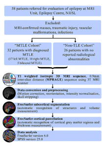
Assessing the brain abnormalities in patients with temporal lobe epilepsy is one area where computerised analysis using FreeSurfer can be applied in a research context. We conducted a study on epilepsy patients at the MRI Unit of the Epilepsy Centre of National Hospital of Sri Lanka utilising FreeSurfer for automated assessment of thickness of the cortical grey matter and volume of subcortical structures. We attempted to identify differences in these parameters in 32 MTLE patients compared with a control group of 26 non-TLE epilepsy patients. We analysed the thickness of grey matter in 34 regions of the cerebral cortex and 25 different volume measurements of brain structures. (Figure 1).
 Figure 1: Study methodology and data processing workflow
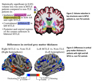
We found significant changes in volume in both temporal and non-temporal lobe structures in patients with TLE in comparison to the controls. Volume was reduced in several regions in the left side of the brain of patients with left-sided MTLE. These regions were the left thalamus, putamen and hippocampus (Figures 2). In patients with right-sided MTLE similar reductions were seen in the right side of the brain in these regions. In some patients the volume of the fourth ventricle of the brain was increased. Total brain volume, volumes of both hippocampi and parts of the corpus callosum were reduced in patients with double-sided MTLE.
Similarly, significant changes were found in the thickness of grey matter of the cortex of the brain in temporal and non-temporal regions in patients with TLE in comparison to the controls. Interestingly, cortical thickness was reduced in certain regions in the right side (fusiform gyrus, banks of superior temporal sulcus) as well as in left side (posterior-cingulate cortex) in patients with right-sided MTLE whereas cortical thickness was increased in certain regions in the right side (entorhinal cortex, medial orbital frontal cortex, insula) in patients with left-sided MTLE. This is a finding that has not been reported in previous studies (Figure 3)
Our study, though limited by the small number of patients, provides a dataset of automatically measured thickness and volume measurements of Sri Lankan MTLE patients which may be useful as the basis for further studies. At the time of conducting the study, we could not identify any prior studies from Sri Lanka which included such measurements using an automated MRI analysis software or manual methods. We also showed that analysis using FreeSurfer can be used as an efficient technique for automated measurement of thickness and volume of brain regions which can be utilised for much more varied purposes in the future. Extensive studies would be required to confirm accuracy of automated FreeSurfer measurements with traditional manual measurements though several existing studies have shown promising comparability.
Ideally normal, healthy volunteers would be used as the control group for comparison, however, due to ethical concerns of subjecting healthy individuals for unnecessary radiological investigations, we opted to use epilepsy patients with non-TLE disorders as the control group, which is a limitation in our current study. Finally, the current study, being a cross-sectional study, cannot distinguish causation from correlation. In other words, from this study we cannot state whether these abnormalities of the brain are a cause for their epilepsy condition or whether they are an effect of it. However, these findings certainly indicate the need for more prospective longitudinal follow-up studies of patients to better understand this debilitating condition.

References
- Fischl B. Freesurfer. NeuroImage. 2012 Aug;62(2):774–81.
